I have a friend who is an artist - he will hold up a flower and say I as an artist can see how beautiful this is, but you as a scientist, oh, take this all apart and it becomes a dull thing. And I think he's kind of nutty… there is also beauty at a smaller dimension, the inner structure … the processes … the beauty that he sees is available to other people [but] a science knowledge only adds to the excitement and mystery and the awe of a flower. - Richard Feynman, The Pleasure of Finding Things Out
Modern microscopic techniques can reveal the beauty of that smaller dimension with mind-boggling clarity. Here too, their allure is made all the greater by understanding the underlying processes, much like how the context, symbolism, or meaning behind a work of visual art can imbue it with a greater sense of significance.
Admittedly, I was once captive to the kind of thinking that plagued Feynman’s friend. About plants especially, I saw them as little more than static parts of my surroundings. A major turning point was in seeing time lapse movies of plant growth and movements, like those by Plants in Motion and BBC’s Life. These films elevated plants for me from unchanging parts of the environment to dynamic and awe-inspiring organisms, ones that simply operate on a different timescale.
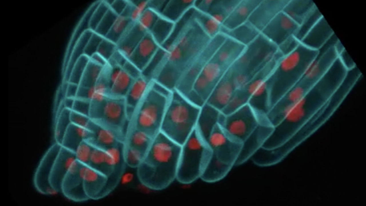
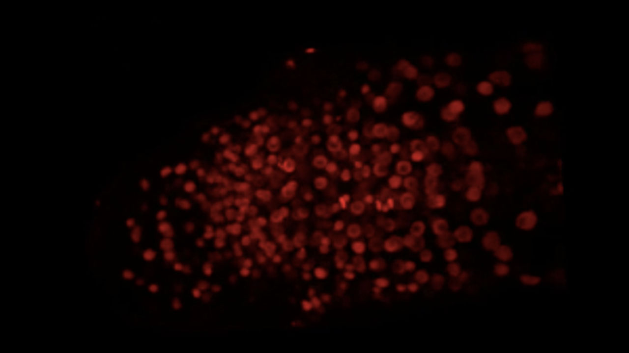
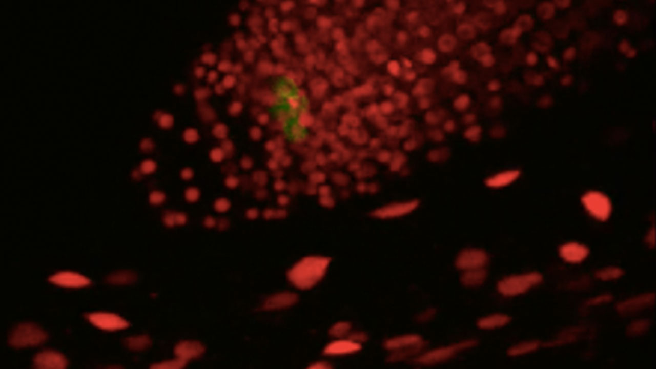
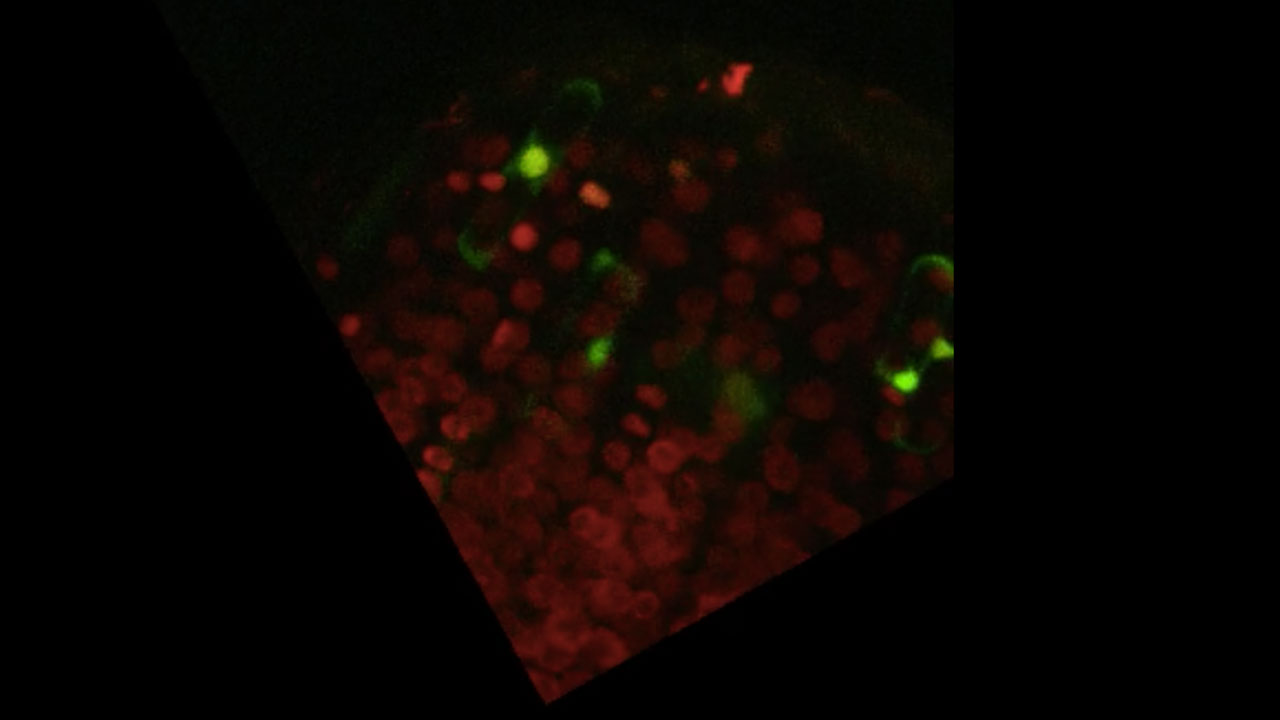
Now, in my PhD project, I use time lapse confocal microscopy to study how plant roots regenerate after severe injury, and I have the privilege of reliving that wonder every day. The films below were made by imaging the entire volume of the root every 13 minutes. Cells’ nuclei are in red.

One Week of Growth - An Arabidopsis root growing for one week. Green marks the Quiescent Center, a small group of cells that sit at the heart of the stem cells in the tip of the growing root.

Successful Regeneration - A root with its tip (including all stem cells) cut away, shown as it regenerates lost tissues and stem cells from the leftover tissue over the course of three days.

Failed Regeneration - Cyan marks the cell membranes in this root, which fails to regenerate after having its tip cut away. Rather than new tissue outgrowth (as in Successful Regeneration), a kind of wound closure takes place. The thin outgrowth at the end of this film is evidence of that cell becoming a root hair, a cell type that is fully differentiated.

Returning Identities - Cells that are resident specifically to the tip can be cut away completely, and be rebuilt from the remaining tissue as the root regenerates. Here we see two cell types, in green and cyan, gradually returning to the root tip after having been cut away. (The bright flashes are short bursts of high laser intensity to try and catch early or faint signs of cells' reappearance.)
.jpg)
Introvert
Introvert - An Arabidopsis root coiled in on itself after growing for one week under a microscope slide.
Is it not a time to break through that dismal convention of the scientific periodicals which orders, however suavely, that only the driest language be used? One would hardly know that these people were making discoveries from the way they have to write them up. - Ithell Colquhoun, Goose of Hermogenes
It only took a half century, but things might finally be moving in a more casual direction. But the parlance of science - both its written and visual language - was once much less dry. Just a half century before that quote, the German biologist Ernst Haeckel released his seminal book, Art Forms in Nature, spanning the divide between art and science. Indeed, early naturalists were often skilled illustrators as well, creating captivating images of newly discovered species that were quite meticulously detailed.
In somewhat more recent times, art and science have had the unfortunate reputation of being dichotomous, and remnants of the false divide carry over to this day, often to the detriment of science communication. When I started graduate school, I was surprised at what little care seemed to go into the visual elements of many professional-level seminars. I was amazed at the rampant, unironic use of Comic Sans and Papyrus fonts in powerpoint talks. Here were highly esteemed scientists at the top of their fields with their presentations dressed up in the typographic equivalent of a bowtie (no disrespect to Mr. Nye).
It was not until my senior year in college that I switched gears to science. As a relative newcomer to the scientific world, I had a personal sense of how inscrutable its content can seem, especially if offered without deliberate care for its presentation. I became quite invested in the visual communication of scientific concepts, both in my published works and in communicating science to lay audiences in general. This has led me to create animated scientific content in partnerships with fellow artist-scientists, such as the pieces below for Science in Real Life, Abstract Zine, and Pondlife.

The Central Dogma of Biology

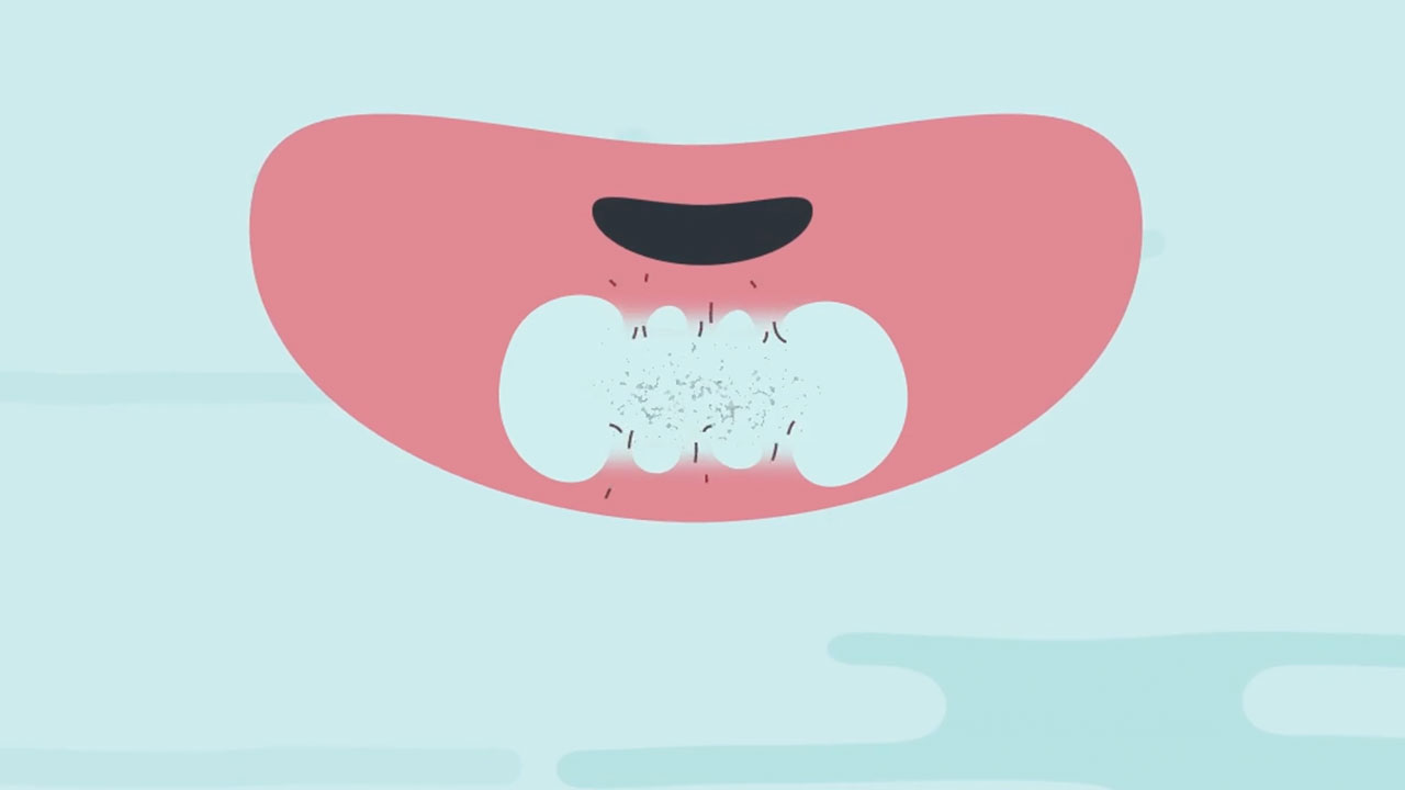
Intestinal Anatomy for Abstract Zine

Symbiogenesis - Every living thing is made up of cells. These cells contain stories. The evolutionary history of how we came to be, from our single cellular ancestors, is one of cooperation between individuals. Here is one of these stories.
About the Author
Ramin is an animator and a biology PhD student in the Birnbaum Lab at New York University. He is godfather to many cats across Brooklyn. You can find more of his work on his personal website.